Previous
| Images
Home | Next
|
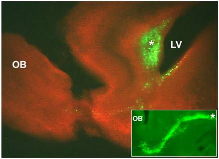
Postnatal
day 2 mouse brain was electroporated with CMV-GFP plasmid into the
anterior lateral ventricle (LV), cut into 250 mm thick slices and
cultured in vitro for 3 days on polycarbonate membrane filters
in serum-free defined media. GFP-positive cells (green)
are seen in the SVZ and a few are migrating toward the olfactory bulb.
The slice was counterstained with propidium iodide (red).
The inset is an example of a slice prepared from another animal and
shows the GFP-labeled cells migrating in the rostral migratory stream.
*, anterior subventricular zone; OB, olfactory bulb.
|
Contact:
parent@umich.edu
Department
of Neurology, University
of Michigan, Ann Arbor, MI
© 2003
All rights reserved.
Web
Design by Michael Serrian/GRAPHIC INSTINCTS