![[ Visit AADR's Website ]](images/banner.jpg)
Objective: Ā Spatiotemporal response of a functional bone-PDL-cementum complex to periodontal disease was evaluated by identifying biochemical and physical changes in the tension and compression sides of the complex.
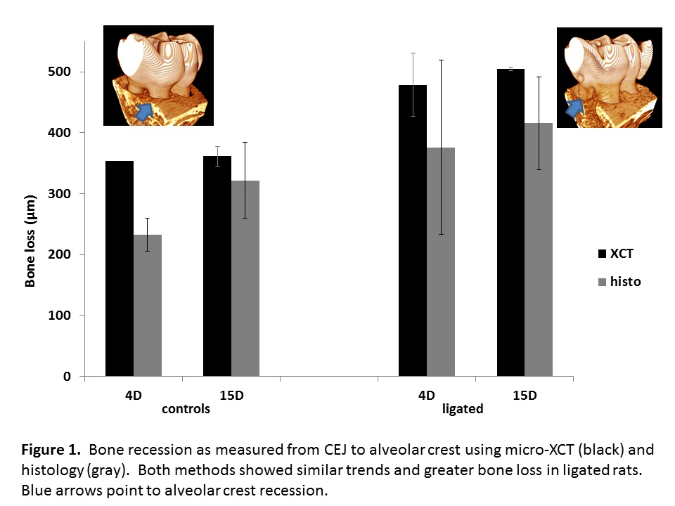
Methods: Ā 4/0 silk ligatures were used to induce periodontitis in 6-week-old male Sprague-Dawley rats for 4 and 15 days; corresponding control rats were flossed (n=3 for each group).Ā Diseased and control maxillae were evaluated using: (1) micro-XCT for PDL-width and alveolar crest recession; (2) histology sections for picrosirius red (PSR), tartrate acid resistant phosphatase (TRAP), and immunofluorescent (IF) labeling of fibronectin (FN), receptor activator of NF-κB ligand (RANKL), and osteopontin (OPN).Ā
Results: ĀIn the naturally prestrained bone-tooth complex (tension in the mesial complex and compression in the distal complex), increased bone recession (Fig. 1) irrespective of tension or compression sides was observed.Ā Bone recession was complemented with the increased presence of TRAP positive cells in the diseased complex.Ā Additionally, increased RANKL and TRAP positive cells in the compression-distal complex compared to the tension-mesial complex (Fig. 2) was observed in diseased specimens.Ā In both controls and diseased, FN expression was identified in secondary cementum.Ā However, in the diseased specimens it was identified on the periphery of cementum resorption pits specifically on the compression side.Ā Higher levels of OPN were identified in control specimens compared to the diseased.Ā Within groups, higher level of OPN was observed in between stratified layers of bone on the tension side of the complex compared to its presence only within the endosteal spaces and around resorption pits on the compression side.
Conclusion: No significant disease related trends were observed between earlier (4D) and later (15D) time points.Ā However, altered biochemical expressions of RANKL, FN and OPN could indicate that disease related effects are more distinct on the compression-distal side of the diseased complex.

Keywords: Biomechanics, Extracellular matrix molecules, Osteoblasts/osteoclasts, Periodontal disease and Resorption