![[ Visit AADR's Website ]](images/banner.jpg)
Infrared Microscopy of Oral Tissues to Detect Foreign Material
Jonathan Prindle, Alfredo Aguirre, Robert E. Baier
Objectives:
1) Demonstrate the ability of infrared microscopy to detect foreign material in oral tissue biopsies using spectrum signature probing of chemical maps. 2) Discern resolution differences between data acquired on germanium substrata in transmission mode, and data acquired on low-e-coated glass in reflection mode. 3) Discern resolution differences with various thicknesses of oral tissue sections.
Methods:
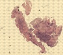
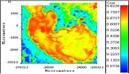
Oral biopsy tissues sections were scanned with the infrared microscope. Each scan created a chemical map that was a collection of individual spectra within a given area. These individual spectra were saved as an integrated chemical map, and the stored file was then probed with stored file reference spectra of various foreign and endogenous substances. Correlation maps between an oral tissue spectrum and reference spectrum showed how highly the chemical nature of the oral tissue matched the chemical nature of the reference spectrum.
Results:
1) Correlation maps between foreign reference spectra, and oral biopsy tissue spectra showed varied and more specific correlations than with endogenous tissue controls, suggesting the presence of specific foreign material in tissues. 2) Positive correlation was found between the thickness of the tissue section and the level of correlation between reference and tissue spectra. 3) Tissues analyzed with infrared beam in transmission through germanium substrata showed improved resolution.
Conclusions:
High correlation between reference and oral biopsy spectra based on similarities in the infrared absorption peaks between the two samples, allowed effective identification of areas where foreign material may be present. Tissue sections of intermediate thickness (2 to 5 micrometers) are needed to prevent loss of resolution. Higher resolution seen on germanium resulted from to germanium's inherent transparency to infrared radiation.
Funding: UB Dental School's Participating Fund for Dental
Education and the Centennial Fund
Keywords: Biomaterials, Microscopy, Oral medicine and Pathology