| Java Not Activated | Java Not Activated | Java Functional |
 |
Blank Area
or message: Image requires a Java enabled browser
|
 |
| KiNG Inactive | KiNG Inactive | KiNG Full Functional |
|
SU Inhibitors Bound To The FGFR 1 Tyrosine Kinase Domain by Larry P. Taylor, Ph. D.
Feedback appreciated; please send comments to: Email: lpt Molecular & Behavioral Neuroscience Institute The University of Michigan Ann Arbor, MI |
My University Home Harris Links Chemistry / Modeling Links
FGF Site: FGF Intro Nomenclature Notes References FGF Sequences FGFR Sequences
Inhibitors Bound To The FGFR 1 Tyrosine Kinase Domain
FGF molecules function by interacting with FGF receptors. These receptors typically contain Ig-like binding domains (with domains D2 and D3 being most involved in FGF-FGF receptor interactions), a short transmembrane spanning domain and a cytoplasmic component that possesses tyrosine kinase activity. An essential component of biological activity is the heparin-mediated dimerization of the FGF receptor.
Since Tyrosine Kinase activity has been observed in many different malignancies and diseases, there is a therapeutic need for specific inhibitors of tyrosine kinase activity.
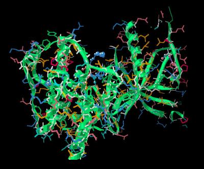
Most FGF receptors contain a tyrosine kinase motif ( Kinemage 1) downstream from the ligand binding domain and membrane spanning region.
Some kinase enzymes contain a WE motif prior to the nucleotide binding loop. The We motif in FGFR 1 is found at Trp-471 and Glu-472. The Glu-472 carboxyl oxygen hydrogen bonds to Thr-552. Kinases containing the WE motif typically hydrogen bond to either a Thr or Ser residue (position 552 in FGFR 1) to stabilize the N-terminal lobe. The WE motif can be considered the boundary between the transmembrane spanning region and the start of the tyrosine kinase domain.
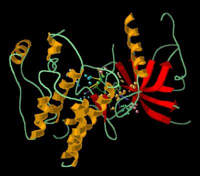
The tyrosine kinase domain is composed of two distinct clusters or lobes separated by a "hinge region" (Tyr-563, Ala-564, Ser-565, Lys-566, Gln-567). The N-terminal region has five anti-parallel beta sheets and one alpha helix. The C-terminal region has two beta strands and seven helices. The two-lobed cluster is a characteristic of tyrosine kinase activity. The "hinge region" accounts for a variety of conformational modes seen between the two distinct lobes of tyrosine kinases.
Unfortunately, in both the unbound and SU-4984 bound crystal structures, the endogenous nucleotide binding loop (Glu-486. Gly-587, Ala-488, Phe-489, Gly-490) is not resolved. This loop is resolved in structure containing bound inhibitor SU-5402.
The catalytic region contains the HRDLAARN sequence commonly found in tyrosine kinases (residues 621-628).
The Inhibitors (Kinemage 2)
SU-4984 (see 1AGW for more discussion)
The ligand binding domain features a hydrophobic cleft (Val-492, Ala-512, Ile-545, Val-461, Ala-564, and Leu-630; that forms a slot for the incoming
oxyindole ring portion of the inhibitor. Residues Leu-484 and Tyr-563 add additional hydrophobic contacts with the inhibitor phenyl ring proximal to the oxyindole. In addition, the inhibitor phenyl ring participates in an oxygen carbonyl to aromatic ring interaction with the carbonyl oxygen of Ala-564. The piperazine ring (most distal from the oxyindole ring) makes a Van der Waal's contact with the highly conserved Gly-567 of the receptor.
The oxyindole portion of the inhibitor makes two hydrogen bonds to the peptide backbone "hinge" region (residues 563-568 between the two distinct lobes of the tyrosine kinase domain of the receptor). These involve the N1 of the ligand oxyindole interacting with the carbonyl oxygen of Glu-562 and the O2 of the oxyindole hydrogen bonding to the amide hydrogen of Ala-564. These same residues are involved in hydrogen bonding to the adenine portion of the endogenous ligand ATP.
SU-5402 (see 1FGI for more discussion)
The SU-5402 inhibitor resides in a cleft of the tyrosine binding region of FGFR 1 that is defined by hydrophobic residues Leu-484, Phe-489, Val-492, Ile-545, Val-561, Tyr-563, Ala-564, and Gly-567. The position of the SU-5402 inhibitor within the binding cleft is anchored by hydrogen bonding between the inhibitor oxyindole ring N1 and the backbone carbonyl of Glu-562, the backbone amide of Ala-564 to the oxyindole ring carbonyl, and the side chain amide of Asn-568 to oxygen of the inhibitor carboxyethyl group.
Of particular significance is the oxy-aromatic interactions of the hydrogen atoms of the plane of Phe-489. All five phenyl group hydrogen atoms are interacting with near-by oxygen atoms: each of the two oxygen atoms of the inhibitor carboxyethyl group, the backbone carbonyl of Arg-627 and the side chain oxygen atoms of Asn-628 and Asp-641. This network of oxy-aromatic interactions and hydrogen bonds most likely explains the preference of SU-5402 for the FGFR 1 receptor. Finally, this area of this receptor's "phenyl loop" is disordered with other inhibitors, but quite well resolved with SU-5402, The SU-5402-FGFR 1 complex is also discussed
The Kinemages
The real-time visualization using KiNG of the structures on this site requires a java-enabled (JRE from Java) browser.
Possible Icons to the left of molecular model image on the download page
| Java Not Activated | Java Not Activated | Java Functional |
|
Blank Area
or message: Image requires a Java enabled browser
|
|
| KiNG Inactive | KiNG Inactive | KiNG Full Functional |
A single click on the KiNG logo will launch the appropriate kinemage.
Kinemage 1: The Tyrosine Kinase Domain
View 1 The Monomer
View 2 WE motif
View 3 Hinge Region
View 3 ATP Catalytic Loop
|
566 K |
|
|
Click On KiNG to see |
Tyrosine Kinase Domain of FGFR 1 |
Kinemage 2: Inhibitor - Receptor Interactions
View 1 The Monomer
View 2 Binding Cleft, "front"
View 3 Binding Cleft, "top"
View 4 H Bonding Interactions
View 5 Oxy-aromatic interactions
|
1.04 M |
|
|
Click On KiNG to see |
Inhibitors Bound To Tyrosine Kinase Domain |
Sequences:
The X-ray resolved residues span residues 464-762 of the human FGFR 1 sequence
For 1AGW:
Unresolved N-terminal: MVAGVSEY
X-ray resolved: ELPEDPRWELPRDRLVLGKPLG
X-ray unresolved: EGAFG
X-ray resolved: QVVLAEAIGLDKDKPNRVTKVAVKM
LKSDATEKDLSDLISEMEMMKMIGKHKNIINLLGACTQDGPLYVIVEYASKGNLREYLQA
RRPPGLEYSYNPSHNPEEQLSSKDLVSCAYQVARGMEYLASKKCIHRDLAARNVLVTEDN
VMKIADFGLARDIHHIDYYKKTTNGRLPVKWMAPEALFDRIYTHQSDVWSFGVLLWEIFT
LGGSPYPGVPVEELFKLLKEGHRMDKPSNCTNELYMMMRDCWHAVPSQRPTFKQLVEDLD
RIVALTS
Unresolved C-terminal: NQE
For 1FGI
Unresolved N-terminal: MVAGVSEY
X-ray resolved: ELPEDPRWELPRDRLVLGKPLGEGAFGQVVLAEAIGLDKDKPNRVTKVAVKM
LKSDATEKDLSDLISEMEMMKMIGKHKNIINLLGACTQDGPLYVIVEYASKGNLREYLQARRPPGLEY
SYNPSHNPEEQLSSKDLVSCAYQVARGMEYLASKKCIHRDLAARNVLVTEDNVMKIADFGLARDIHHI
DYYKKTTNGRLPVKWMAPEALFDRIYTHQSDVWSFGVLLWEIFTLGGSPYPGVPVEELFKLLKEGHRM
DKPSNCTNELYMMMRDCWHAVPSQRPTFKQLVEDLDRIVALTS
Unresolved C-terminal: NQE
Sources:
Residues 464-762 of human FGFR 1 was isolated from transfected Spodoptera frugiperda insect cells; structural coordinates were taken from the Brookhaven Database files 1AWG (SU 4984) 1FGI (SU 5402). The individual monomers were extracted with SYBYL and then aligned with the "magic fit" option of Deep View.
The engineered sequence contained three residues that were different from human:
Cys-488 and 584 were changed to Ser to prevent disulfide oligiomerization.
Leu-457 was changed to Val to create an aNcol cloning site.
FGF Site: FGF Intro Nomenclature Notes References FGF Sequences FGFR Sequences
My University Home Harris Links Chemistry / Modeling Links
Copyright 2006-2020 by Larry P. Taylor
Molecular & Behavioral Neuroscience Institute
The University of Michigan
All Rights Reserved
Supported by the Pritzker Neuropsychiatric Disorders Research Consortium, and by NIH Grant 5 P01 MH42251, Conte Center Grant #L99MH60398, RO1 DA13386 and the Office of Naval Research (ONR) N00014-02-1-0879 to Huda Akil & Stanley J. Watson. at the Molecular & Behavioral Neuroscience Institute.