| Java Not Activated | Java Not Activated | Java Functional |
 |
Blank Area
or message: Image requires a Java enabled browser
|
 |
| KiNG Inactive | KiNG Inactive | KiNG Full Functional |
|
Human Fibroblast Growth Factor 4 by Larry P. Taylor, Ph. D.
Feedback appreciated; please send comments to: Email: lpt Molecular & Behavioral Neuroscience Institute The University of Michigan Ann Arbor, MI |
My University Home Harris Links Chemistry / Modeling Links
FGF Site: FGF Intro Nomenclature Notes References FGF Sequences FGFR Sequences
Human Fibroblast Growth Factor 4
Growth factors represent a family of at least 18 proteins with a beta trefoil (three-fold repeat of a four-stranded sheet assembly without extended alpha helix strands) motif associated with cell proliferation and differentiation. They are a prime component of angiogenesis associated with organogenesis, tumor growth, and wound healing.
FGF molecules function by interacting with FGF receptors. These receptors typically contain Ig-like binding domains (with domains D2 and D3 being most involved in FGF-FGF receptor interactions), a short transmembrane spanning domain and a cytoplasmic component that possesses tyrosine kinase activity. An essential component of biological activity is the heparin-mediated dimerization of the FGF receptor.
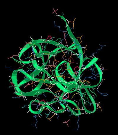
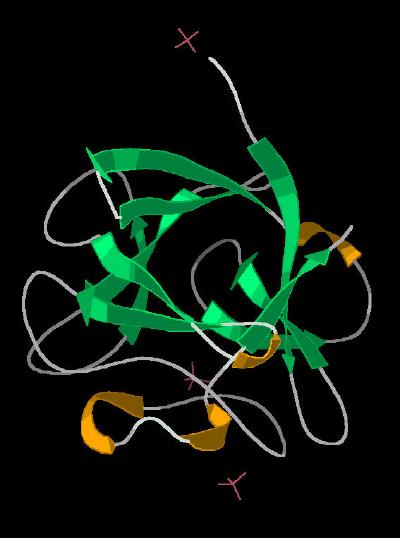
The FGF 4 molecule is shown with ribbon rendering in Kinemage 1. The cartoon rendering is displayed in
Kinemage 2.
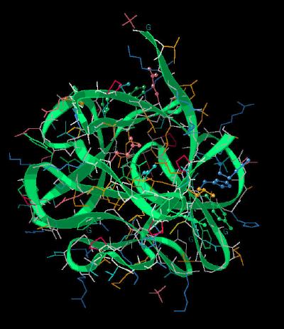
Kinemage 3 highlights the FGF residues involved in ligand-receptor interactions (based on a structural overlay of FGF 4 upon an FGF 2 molecule binding to the FGFR 1 receptor.
Differences between FGF 4 and FGF 1 & FGF 2 occur primarily at beta sheets 1 and 2, 3 and 4, and 9 and 10. These loops interact with the ligand binding domain of FGF receptors and are most likely the determining factor in FGF 4's binding profile.
Residues implicated in major hydrophobic contact roles with the D2 Domain of the receptor include conservative residues Tyr-87, Tyr-166, and Leu-203. FGF 4 residues His-95 and Arg-205 lessen hydrophobic interactions between ligand and receptor, especially compared to FGF 2 comparable residues Phe-40 and Met-151.
Conserved residue Asn-167 forms a critical H-bond to the invariant D2-D3 Linker Arg residue.
Ligand-receptor interactions at D3 center around contact between invariant residue Glu-159 and receptor invariant residue Other important residues include Glu-117, Ser-119, Val-121, Phe-129, Phe-136, and Phe-151.
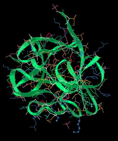
Kinemage 4 highlights residues that interact with heparin. They are Asn-89, Lys-183, and Lys-188.
FGF 4 has approximately 30% sequence identify of the FGF 1 & 2 sequences. It is unique in that the molecule is the only FGF molecule with a signal sequence preceding the trefoil structure of the growth factor, FGF 4 shows a binding preference for the IIIc splice variant of FGFR 1-3, with little binding to either the IIIb variant of FGFR 1-3 or FGFR 4. A structural overlay of FGF 4 compared to FGF 1 and FGF 2 is shown in
Kinemage 5.
The Kinemages:
The real-time visualization using KiNG of the structures on this site requires a java-enabled (JRE from Java) browser.
Possible Icons to the left of molecular model image on the download page
| Java Not Activated | Java Not Activated | Java Functional |
|
Blank Area
or message: Image requires a Java enabled browser
|
|
| KiNG Inactive | KiNG Inactive | KiNG Full Functional |
A single click on the KiNG logo will launch the appropriate kinemage.
Kinemage 1: FGF 4 Ribbon Rendering
|
211 K |
|
|
Click on KiNG to see |
FGF 4 Ribbon |
Kinemage 2: FGF 4 Cartoon Rendering
|
170 K |
|
|
Click on KiNG to see |
FGF 4 Cartoon |
Kinemage 3: Receptor Binding Residues
|
180 K |
|
|
Click on KiNG to see |
Receptor Binding Residues |
Kinemage 4: Heparin Binding Residues
|
180 K |
|
|
Click on KiNG to see |
Heparin Binding Residues |
Kinemage 5 : Backbone Overlays
View 1 The Overlays
View 2 Sheet 1 & 2
View 3 Sheet 3 & 4
View 4 Sheet 9 & 10
|
507 K |
|
|
Click on KiNG to see |
Backbone Overlays |
Sequence: (corresponding to human FGF 4 (79-206 )
GIKRLRRLYCNVGIGFHLQALPDGRIGGAHADTRDSLLELSPVERGVVSIFGVASRFFVAMSSKGKLYGSPFFTDECTFK
EILLPNNYNAYESYKYPGMFIALGKNGKTKKGNRVSPTMKVTHFLPRL
Source:
The appropriate DNA sequence was subcloned into the pET-15b bacterial expression vector and transformed in E coli. The structural coordinates were taken from Brookhaven Database File 1IJT.
FGF Site: FGF Intro Nomenclature Notes References FGF Sequences FGFR Sequences
My University Home Harris Links Chemistry / Modeling Links
Copyright 2005-2020 by Larry P. Taylor
Molecular & Behavioral Neuroscience Institute
University of Michigan
All Rights Reserved
Supported by the Pritzker Neuropsychiatric Disorders Research Consortium, and by NIH Grant 5 P01 MH42251, Conte Center Grant #L99MH60398, RO1 DA13386 and the Office of Naval Research (ONR) N00014-02-1-0879 to Huda Akil & Stanley J. Watson. at the Molecular & Behavioral Neuroscience Institute.