
Sequential axial T2 images through the cerebellum from inferior to superior (left to right) show decreased edema throughout the cerebellar hemispheres with improved definition of the cerebellar folia. There is also decreased effacement of the 4th ventricle (top right image vs bottom right image)
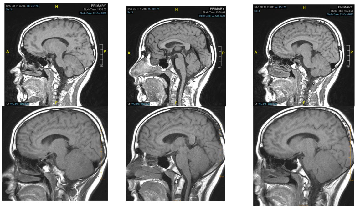
Sagittal T1 images from left to right similarly demonstrate markedly improved definition of the cerebellar folia with decreased effacement of the 4th ventricle (top middle vs bottom middle image)
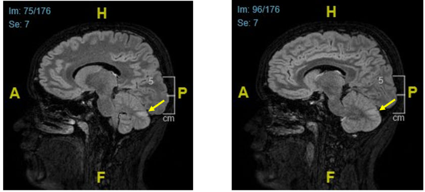
Sagittal T2 FLAIR through the right and left cerebellar folia show some residual edema (arrows).