
Cardiac Tissue Engineering
Robert G. Dennis, Ph.D.

|
|
Cardiac Tissue Engineering Robert G. Dennis, Ph.D. |
|
Objective: To engineer functional mammalian cardiac muscle constructs in vitro.
Experimental Approach:
Our work in this area is still very preliminary,
and I am seeking funding for additional research. Video clips of
the self organizing cardiac tissue are provided below.
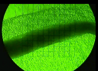 |
The image to the left is a still frame
from a video of a monolayer of cardiac myocytes, self-organizing into a
culindrical tissue structure in culture (video taped on January 1, 2002).
The monolayer of cells is actually composed of both primary mammalian cardiac
myocytes and fibroblasts. Note that as the monolayer contracts spontaneously
(in real time), the cell monolayer peels away from the substrate material
(top of frame) a small amount with each contraction. Under the correct
culture conditions, the self-organization of the cardiac tissue is visible
with the naked eye (no magnification is required)
The square grid pattern is 1mm x 1mm in size, the smaller squares being 100 microns on each edge. To see
the full length video, click this link (~17MB)
(Photograph and video by R.G. Dennis 1/1/2002) |
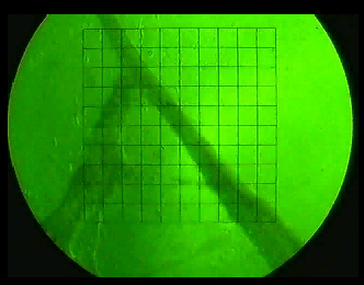 |
The image to the left is a still frame
from a video of a randomly-organized cardiac muscle tissue mass.
Without anchor points to define the long axis of the self-organizing tissue
mass, the cardiac tissue will self-organize into a random web of spontaneously
contracting tissue.
The square grid pattern is 1mm x 1mm in size, the smaller squares being 100 microns on each edge. To see
the full length video, click this link (~2.9MB)
(Photograph and video by R.G. Dennis 1/1/2002) |
Bob's Home Page Current Research Muscle Mechanics Lab (U of M) Biomechatronics Group @ MIT